Clinical
Case Report
Objective Soft Tissue Assessment Supports Optimal Surgical Timing When Clinical Signs Are Inconclusive

Preliminary data shared prior to publication with allowance by Prof. Dr. med. R. Sellei, Chief Surgeon and Head of the Department of Trauma Surgery and Orthopaedic Surgery, Sana Klinikum Offenbach, Germany.
Objective
Case Presentation
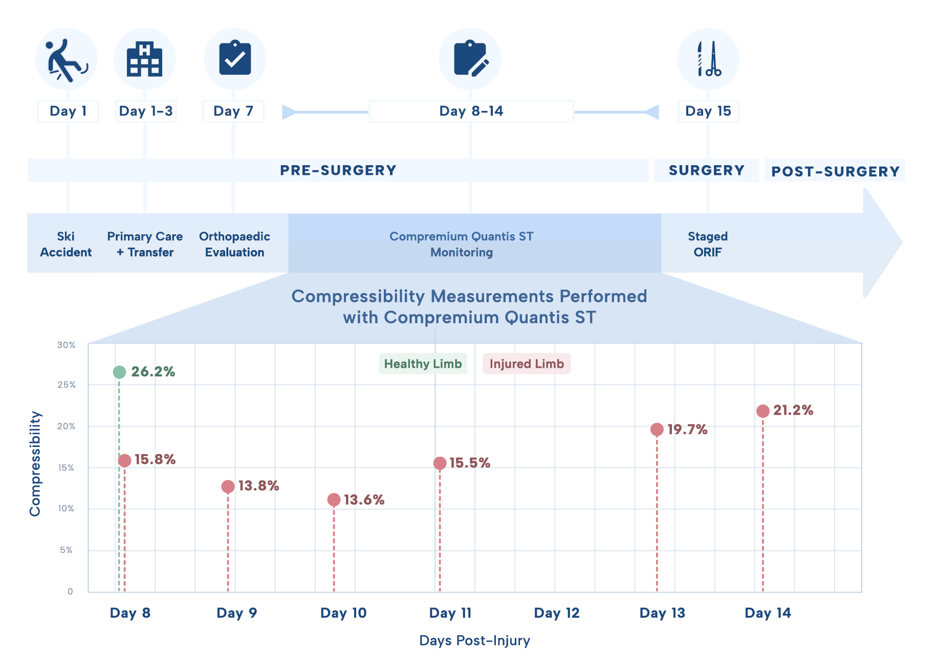
A 57-year-old male sustained a Schatzker VI left tibial plateau fracture following a ski accident, complicated by an erysipelas.
Day 1:
Initial management took place in Austria on the day of the accident, where the injury was primarily stabilized with an external fixator.
Day 3:
The patient was transferred for further care, and the pin-site infection was treated with intravenous antibiotics.
Clinical assessment:
Despite a positive wrinkle sign, persistent soft tissue swelling raised concern that the subfascial compartment had not yet recovered.
Day 4-5:
The decision point was timing of definitive ORIF. To objectify soft-tissue readiness, serial non-invasive compressibility measurements were performed using Compremium Quantis ST on the injured limb, referenced against the uninjured contralateral side.
Solution
Compressibility Measurements
Day 8–11:
Epifascial signs are favorable (positive wrinkle sign, regressing erysipelas)
Compressibility values at ~16%, significantly below the contralateral baseline of 26.2%.
The soft tissue was not yet fit for ORIF, with elevated risk of dehiscence and infection.
From Day 13:
compressibility approached 20%, consistent with adequate soft-tissue recovery for surgery.
Result
Outcome & Conclusion
Definitive fixation was deferred until objective soft-tissue recovery, avoiding intervention while the compartment remained congested.
Once values approached the contralateral baseline, surgical timing was confirmed on quantitative grounds rather than surface signs alone.
The patient underwent a 360° surgical procedure in three stages:
Day 15: ORIF via dorsomedial approach (LCP 3.5 mm) .
Day 21: ORIF via anteromedial approach with defect bridging (Cronos block).
1 Month After Injury: Lateral ORIF with re-osteosynthesis of the tuberositas tibiae.
Outcome: Uneventful wound healing, no infection, no revision.
Compremium Quantis ST delivered a repeated, quantitative, non-invasive measurement to support clinical decision making at the bedside.
ORIF on a compromised soft-tissue was avoided, minimizing potential risks of infection and subsequent revision.
Surgical timing was driven by objective subfascial data, not surface signs alone; removing uncertainty in soft-tissue judgment.
CE-approved intended use
The CPMX1 Software is intended for real-time and intermittent measurement and monitoring of relative compartment compressibility.
FDA-cleared intended use
The Compartmental Compressibility Monitoring System (CPM#1) is intended for real-time and intermittent monitoring of relative compartment compressibility. The relative compartment compressibility (CP Value) is not meant for trend analysis. 510(k) Number: K223509.


